How virtual thin sections are created
Many samples in the Virtual Microscope were created from thin sections produced by the Open University laboratory. However, an increasing number of the thin sections on the website were created by labs at other institutions. Some of our thin sections are almost 100 years old!
The Open University has a well-equipped sample preparation laboratory that produces high quality thin sections up to 50 x 75 mm in size. Typically, terrestrial rocks are cut using a trim saw, ground flat (lapped), and then mounted on glass slides before being ground until they are 30 microns (0.03mm) thick and finished by hand. If samples are to be used for reflected light viewing (for ore microscopy) or micro-analysis (e.g. electron microprobe), then the final stage of preparation involves aluminium oxide or diamond polishing. Extraterrestrial samples follow a similar preparation procedure, although a wafer saw is used to minimise sample loss and special precautions are employed to avoid contamination.
Thin sections for the Virtual Microscope have to be very high quality and ultra-clean, but the perfect section does not exist. Unlike a conventional thin section, it is possible for a virtual microscope thin section to be seen by thousands of people, so a lot of effort goes into producing quality sections. In the course of preparing samples for the Virtual Microscope we have seen quite a few things that are artefacts and contamination.
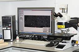
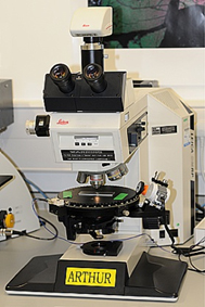
We currently scan thin sections using a Leica DM 2500P Pol microscope (with transmitted and reflected light capability) and a Leica fast acquisition digital microscope camera (DFC400). Images are matched and stitched using Leica Power Mosaic software and a Leica PC computer. The system produces seamless high-resolution mosaics that can reach up to 4 GB in size.
As part of our response to the coronavirus situation, we collaborated with universities who acquired digital scans of their teaching samples and sent them to us for processing and upload. These virtual samples were delivered as Collections on the Virtual Microscope website for the universities’ own students, but they were also made freely available in line with the aims of the Virtual Microscope project.
The 'raw' resources from which the specimen is created are combined in a folder structure with the HTML5 viewer software on the server. Each Virtual Microscope thin section is presented in an iframe by the Drupal management system, allowing users to study the thin sections at the same resolution at which they were created. The HTML5 Virtual Microscope application will work across most platforms, including Windows, Mac OS and Linux, and will also work on the iPad and android tablets.
Further information
If you would like to know more about the features you see in the Virtual Microscope, or how geologists identify and classify rocks and minerals, check out these resources.